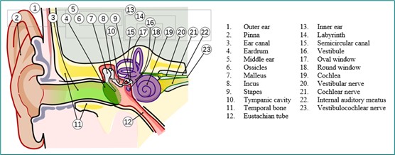
Figure 2.4. The cochlear nerve is located in the inner ear.
Source: https://commons.wikimedia.org/wiki/File:Anatomy_of_the_Human_Ear_1_Intl.svgShort alt text: An anatomical drawing of an ear with parts labeled.
Long alt text: The drawing displays the components of the ear from left to right: pinna, outer ear, ear canal, temporal bone, eardrum, middle ear, ossicles, malleus, incus, tympanic cavity, stapes, inner ear, semicircular canal, labyrinth, oval window, vestibule, round window, eustachian tube, cochlea, vestibular nerve, cochlear nerve, internal auditory meatus, and vestibulocochlear nerve. The cochlear nerve is one of the innermost parts, extending further inward from the cochlea at the front of the inner ear.
Editor’s Tip
Notice how we only describe positioning of the cochlear nerve, as that was mentioned in the caption and is the focus of the image. The caption and surrounding text will indicate if the full diagram needs to be described or only part.


